
Lactate Metabolism Plays an Important Role in Mitochondrial Diseases
January 11, 2023Lactate, long considered to be a waste product of energy production, has been found to fuel the metabolism of patients…
Mitochondrial diseases affect approximately 1 in every 5,000 newborns, making them some of the most common genetic diseases. How these diseases present varies greatly, ranging from early fatality to progressive, adult-onset symptoms. This makes diagnosis and prognosis difficult, and even with proper identification, there are currently no effective treatments for this class of diseases. The best chance for developing treatments lies in expanding our understanding of mitochondrial behavior. This is particularly true within the context of the whole cell and organism because mitochondria are considered metabolic “hotspots” and are well-integrated into a large number of biochemical pathways.
Our research focuses on how mitochondria are embedded in normal cellular function. Improving our understanding will help us identify new insights into mitochondrial diseases and allow us
to develop clinical tools and therapeutic options.
Mitochondrial Dynamics and Metabolic Homeostasis
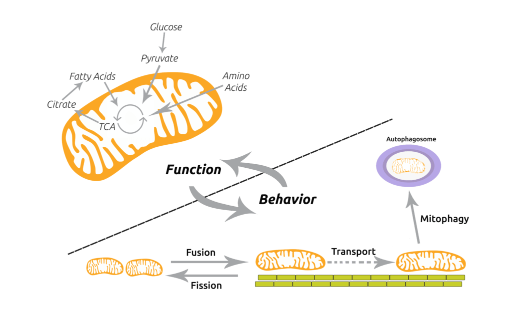
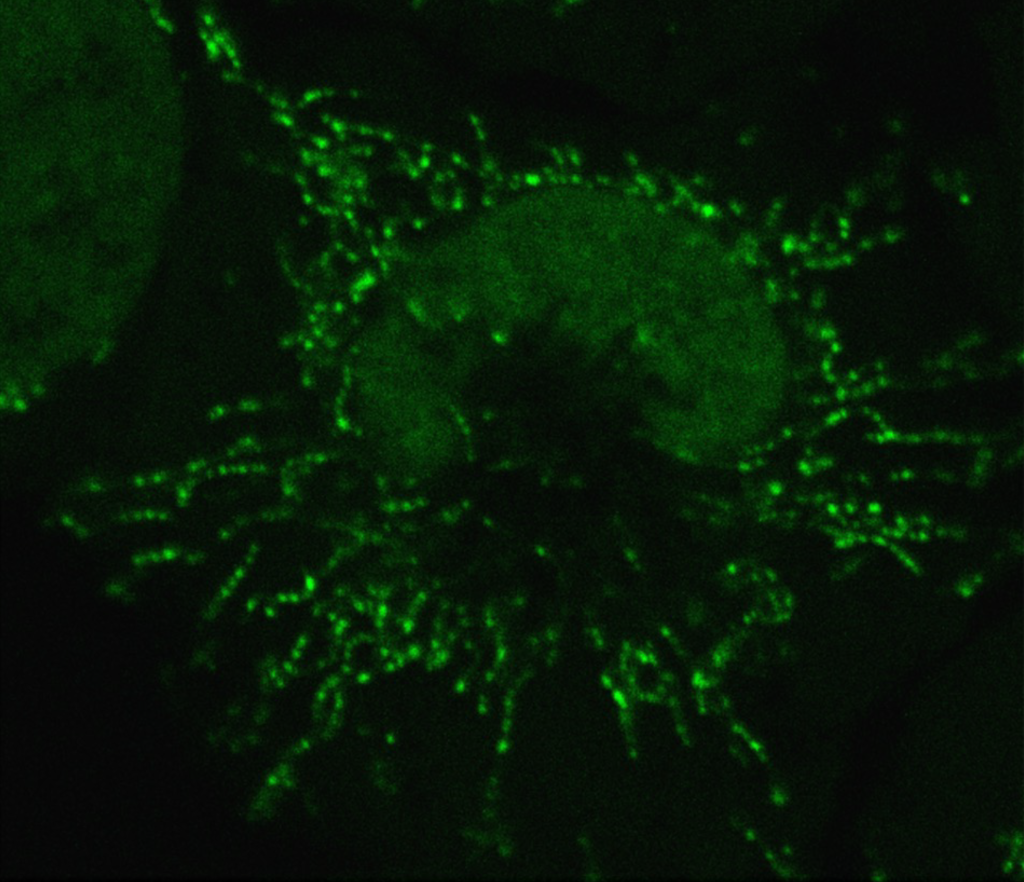
Mitochondria play a central role in cellular metabolism and produce ATP from numerous fuel sources and participate in many biochemical pathways, such as amino acid metabolism, Fe-S cluster assembly and calcium handling. Mitochondria also exhibit interesting macroscopic behaviors, including fusion (joining of two organelles into one), fission (division), active transport along cytoskeletal elements and mitophagy (targeted destruction). Together, these behaviors are known as mitochondrial dynamics.
On the surface, mitochondrial behaviors are functionally independent from their biochemical roles and mediated by distinct proteins. However, recent data from our lab and others suggest metabolism influences mitochondrial behavior and vice versa. For instance, culture conditions that enhance the oxidative phosphorylation activity of mitochondria serve to increase fusion rates (Mishra et al., Cell Metabolism 2014) and increase mitophagy rates (Melser et al., Cell Metabolism 2013). Conversely, genetic ablation of mitochondrial fusion results in severe OXPHOS defects (Chen et al., JBC 2005).
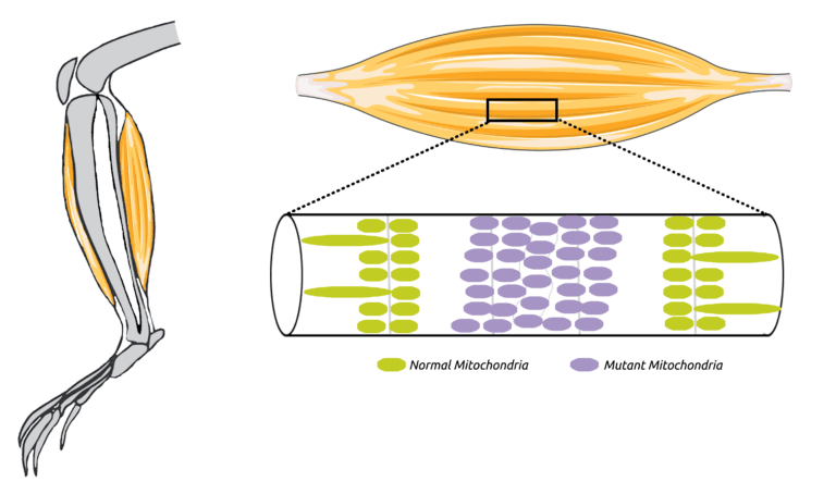
Muscle dysfunction and weakness are common symptoms of mitochondrial diseases, caused by mutations within the mitochondrial genome (mtDNA). In comparison with other myopathies, mitochondrial myopathies have some distinct features. In particular, individual muscle fibers are affected in a regional manner with affected segments surrounded by normal regions of the fiber (see right). Affected regions are classified by a loss of mitochondrial activity and an accumulation of mutant mitochondria. Interestingly, the mutation does not spread beyond the affected region, which suggests that mechanisms are in place to restrict the defect from disrupting the whole fiber.
We have recently developed methods to quantify mitochondrial regionalization within muscle fibers (Mishra et al., Cell Metabolism 2015). Satellite cells are muscle-resident stem cells that incorporate into existing myofibers. Using a satellite-cell specific promoter, we have stochastically activated individual myonuclei in live animals to label mitochondria in their immediate vicinity (see below). The spread of the mitochondrial signal provides a relative measure of the extent of regionalization, which we term a mitochondrial “domain”. We have found the type of muscle fiber, as well as the extent of mitochondrial fusion occurring in that fiber, determines domain size. Indeed, mitochondrial fusion appears to promote spreading of the activated signal, thereby extending the length of the domain.
We therefore hypothesize that fusion rates play a role in determining the progression of mitochondrial disease. Using mouse models of disease in combination with conditional knockout alleles for pro-fusion proteins (mitofusin 1 and 2), we will test this hypothesis. Data from these studies will suggest whether modulation of organellar dynamics may be beneficial in the treatment of mitochondrial diseases.
Prashant Mishra received his undergraduate degree in biochemical sciences from Harvard University. He received his M.D. and Ph.D. degrees from the University of Texas Southwestern Medical Center, where he studied the role of scaffolding in signaling pathways with Dr. Rama Ranganathan. As a fellow of the Jane Coffin Childs Memorial Fund, he conducted postdoctoral work on regulation of mitochondrial behavior in the laboratory of Dr. David Chan at the California Institute of Technology.
In 2015, he joined the faculty of the Children’s Medical Center Research Institute at UT Southwestern.
Wang, X., and Mishra P. (2023). Fusion of dysfunction muscle stem cells with myofibers induces sarcopenia in mice. bioRxiv. 10.1101/2023.01.20.524967. (PubMed)
Chen, Z., Bordieanu, B., Kesavan, R., Lesner, N.P., Venigalla, S.S.K., Shelton, S.D., DeBerardinis, R.J., and Mishra, P. (2022). Lactate metabolism is essential in early-onset mitochondrial myopathy. Sci. Adv. 9(1):eadd3216. (PubMed)
Garg, A., Keng, W.T., Chen, Z., Sathe, A.A., Xing, C., Kailasam, P.D., Shao, Y., Lesner, N.P., Llamas, C.B., Agarwal, A.K., and Mishra, P. (2022). Autosomal recessive progeroid syndrome due to homozygosity for a TOMM7 variant. J Clin Invest. 132, e156864. (PubMed)
Lesner, N.P., Wang, X., Chen, Z., Frank, A., Menezes, C., House, S., Shelton, S.D., Lemoff, A., McFadden, D.G., Wanaspura, J., DeBerardinis, R.J., and Mishra, P. (2022). Differential requirements for mitochondrial electron transport chain components in the adult murine liver. Elife. 11, e80919. (PubMed)
Wang, X., Jia, Y., Zhao, J., Lesner, N.P., Menezes, C.J., Shelton, S.D., Venigalla, S.S.K., Xu, J., Cai, C., and Mishra, P. (2022). A Mitofusin 2 – Hif1α axis sets a maturation checkpoint in regenerating skeletal muscle. J Clin Invest. 2, 155-169. (PubMed)
Wang, X., Shelton, S.D., Bordieanu, B., Frank, A.R., Yi, Y., Venigalla, S.S.K., Gu, Z., Lenser, N.P., Glogauer, M., Chandel, N.S., Zhao, H., Zhao, Z., McFadden, D.G., and Mishra, P. (2022). Scinderin promotes fusion of electron transport chain dysfunctional muscle stem cells with myofibers. Nat Aging. 2, 155-169. (PubMed)
Lesner, N.P., Gokhale, A., Kota, K., DeBerardinis, R.J., and Mishra, P. (2020). α-ketobutyrate links alterations in cystine metabolism to glucose oxidation in mtDNA mutant cells. Metab Eng. 60, 157-167. (PubMed)
Mishra, P. (2016). Interfaces between mitochondrial dynamics and disease. Cell Calcium 60, 190—198. (PubMed)
Mishra, P., and Chan, D.C. (2016). Metabolic regulation of mitochondrial dynamics. J Cell Biol 212, 379—387. (PubMed)
Mishra, P.*, Varuzhanyan, G.*, Pham, A.H., and Chan, D.C. (2015). Mitochondrial dynamics is a distinguishing feature of skeletal muscle fiber types and regulates organellar compartmentalization. Cell Metab 22, 1033—1044. (PubMed) *co-first author
Mishra, P., and Chan, D.C. (2014). Mitochondrial dynamics and inheritance during cell division, development and disease. Nat Rev Mol Cell Biol 15, 634—646. (PubMed)
Lactate, long considered to be a waste product of energy production, has been found to fuel the metabolism of patients…
Recovery time for muscle sprains, strains, and other soft tissue injuries could be shortened thanks to the discovery of a…
Within our cells, mitochondria are responsible for converting food into energy. This works by taking electrons from the nutrients we…
Ph.D. Student (2017-2021)