A team of scientists at the Children’s Research Institute at UT Southwestern (CRI) has become the first to use a tissue-clearing technique to localize a rare stem cell population, in the process cracking open a black box containing detailed information about where blood-forming stem cells are located and how they are maintained. The findings, published in Nature, provide a significant advance toward understanding the microenvironment in which stem cells reside within the bone marrow.
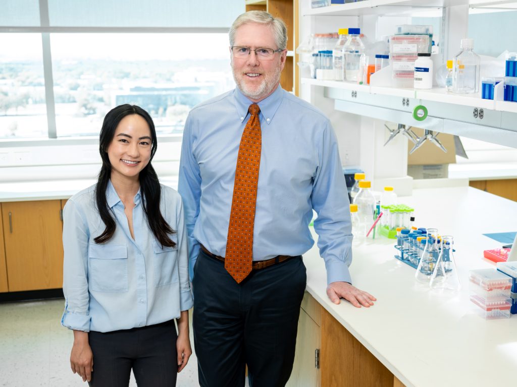
“The bone marrow and blood-forming stem cells are like a haystack with needles inside. Researchers in the past have been able to find a few stem cells, but they’ve only seen a small percentage of the stem cells that are there, so there has been some controversy about where exactly they’re located,” said Dr. Sean Morrison, CRI Director and Mary McDermott Cook Chair in Pediatric Genetics at UT Southwestern Medical Center. “We developed a technique that allows us to digitally reconstruct the entire haystack and see all the needles — all the blood-forming stem cells that are present in the bone marrow — and to know exactly where they are and how far they are from every other cell type.”
The CRI team began by identifying a genetic marker that is almost exclusively expressed in blood-forming stem cells. They took green fluorescent protein from jellyfish and inserted it into the genetic marker to be able to visually identify the stem cells. The fluorescent protein makes the stem cells glow green within the bone marrow.
“Using a tissue-clearing technique that makes the bone and bone marrow see-through, and employing a high-resolution, confocal microscope to scan the entire bone marrow compartment, we were able to image large segments of bone marrow to locate every blood-forming stem cell and its relation to other cells,” said Dr. Melih Acar, an Assistant Instructor at CRI and the paper’s first author. The team’s work yielded new findings and confirmed others: blood-forming stem cells tend to be clustered in the center of the bone marrow, not closer to bone surfaces as some had previously thought; blood-forming stem cells are indeed associated with sinusoidal blood vessels; and there are no spatially distinct niches for dividing and non-dividing blood-forming stem cells.
“With this improved understanding of the microenvironment and mechanisms that maintain blood-forming stem cells, we are closer to being able to replicate the environment for blood-forming stem cells in culture,” said Dr. Morrison, who is also a CPRIT Scholar in Cancer Research and a Howard Hughes Medical Institute Investigator. “That achievement would significantly improve the safety and effectiveness of bone marrow transplants and potentially save thousands of additional lives each year.”
Other CRI researchers involved in the research were Dr. Kiranmai Kocherlakota, Dr. Malea Murphy and Dr. James Peyer. The work was supported by the National Institutes of Health, the Cancer and Prevention Research Institute of Texas, and donors to the Children’s Medical Center Foundation.
About CRI
Children’s Medical Center Research Institute at UT Southwestern (CRI) is a joint venture established in 2011 to build upon the comprehensive clinical expertise of Children’s Health System of Texas and the internationally recognized scientific excellence of UT Southwestern Medical Center. CRI’s mission is to perform transformative biomedical research to better understand the biological basis of disease, seeking breakthroughs that can change scientific fields and yield new strategies for treating disease. Located in Dallas, Texas, CRI is creating interdisciplinary groups of exceptional scientists and physicians to pursue research at the interface of regenerative medicine, cancer biology and metabolism, fields that hold uncommon potential for advancing science and medicine. More information about CRI is available on its website: cri.utsw.edu.