Scientists in the McBrayer lab in Children’s Medical Center Research Institute at UT Southwestern (CRI) have developed a technique that, for the first time, allows a deadly type of brain tumor to exist outside the body and be studied in the laboratory. The new model, published in Neuro-Oncology, offers researchers the opportunity to study how the microenvironment influences brain tumor biology as well as test promising new therapies for glioma.
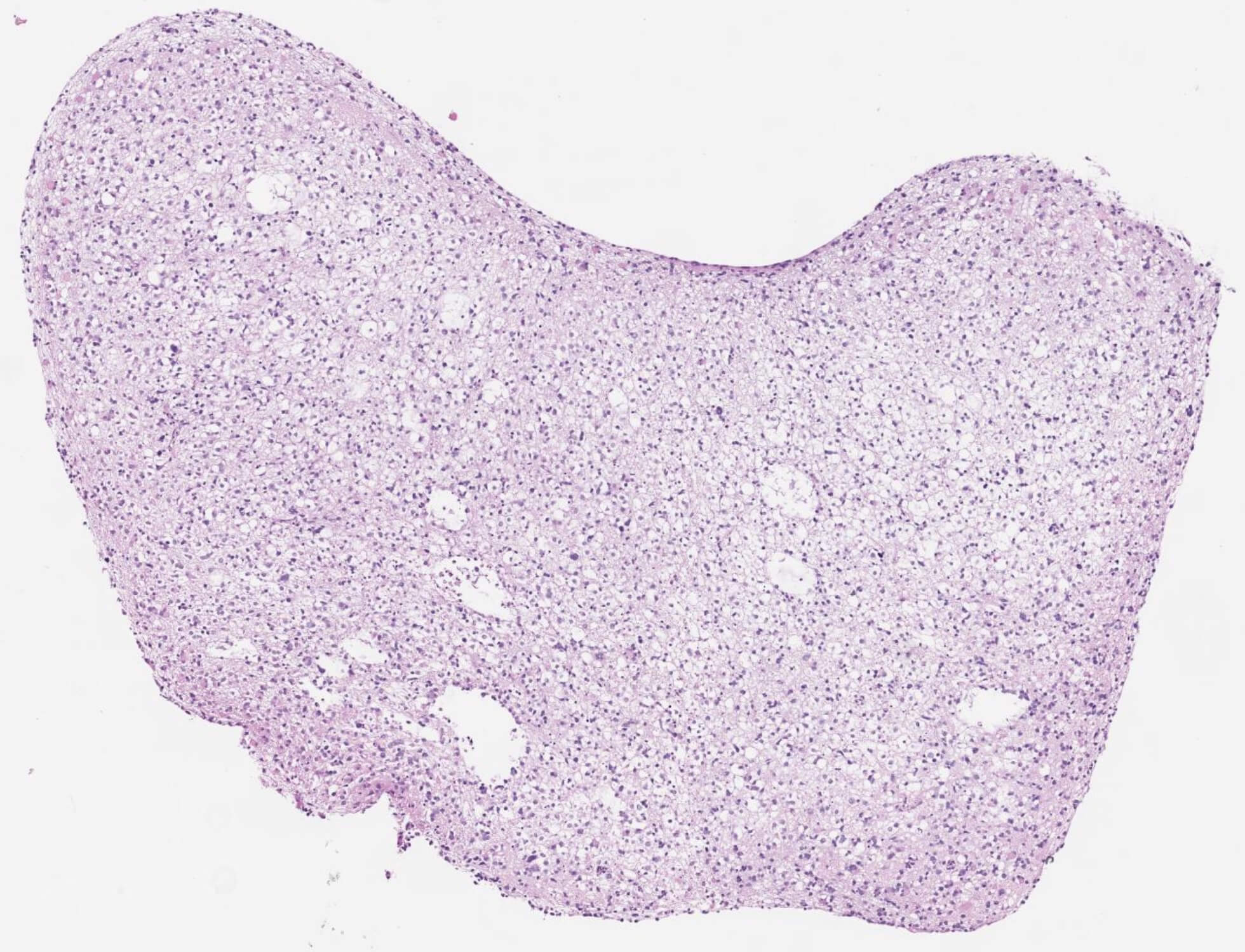
H&E stain of low-grade glioma organoid.
Low-grade gliomas (LGGs) are among the most common malignant brain tumors affecting young adults. Resistant to chemotherapy and difficult to treat, they invariably progress to high-gliomas (HGGs), which carry a 5-year survival rate under 5%. The inability to accurately model LGG is a major factor contributing to the lack of progress in developing effective new brain tumor therapies. In contrast to HGGs, LGGs have not been previously grown in the lab. To address this problem, Dr. McBrayer and his collaborators identified key metabolic conditions that allowed LGG tumor samples to be successfully propagated in the laboratory setting.
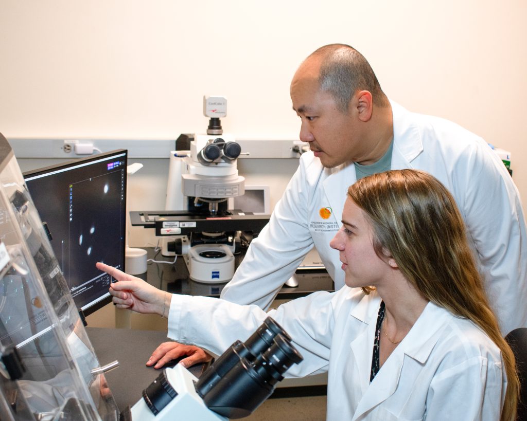
“We worked with our partners in the Department of Neurological Surgery to obtain tumor tissue from patients undergoing brain tumor resections. We then used that tissue to create miniature versions of these tumors, called organoids. This new model allows us to dig into every aspect of any molecular or chemical weakness that might be a target for new cancer drugs and study the diverse cell populations that make up the tumor,” said Samuel McBrayer, Ph.D., Assistant Professor at CRI.
The organoids – which are about the width of a pencil tip – displayed visual, genetic, and immunological characteristics nearly identical to the tumors they are taken from. They can be kept alive for up to six months in the lab or frozen and reanimated, allowing them to be stored for future use without damaging the tissue. Eventually, these new organoids may allow scientists to test drugs for patients as they undergo cancer treatment, a step toward highly personalized brain tumor care.
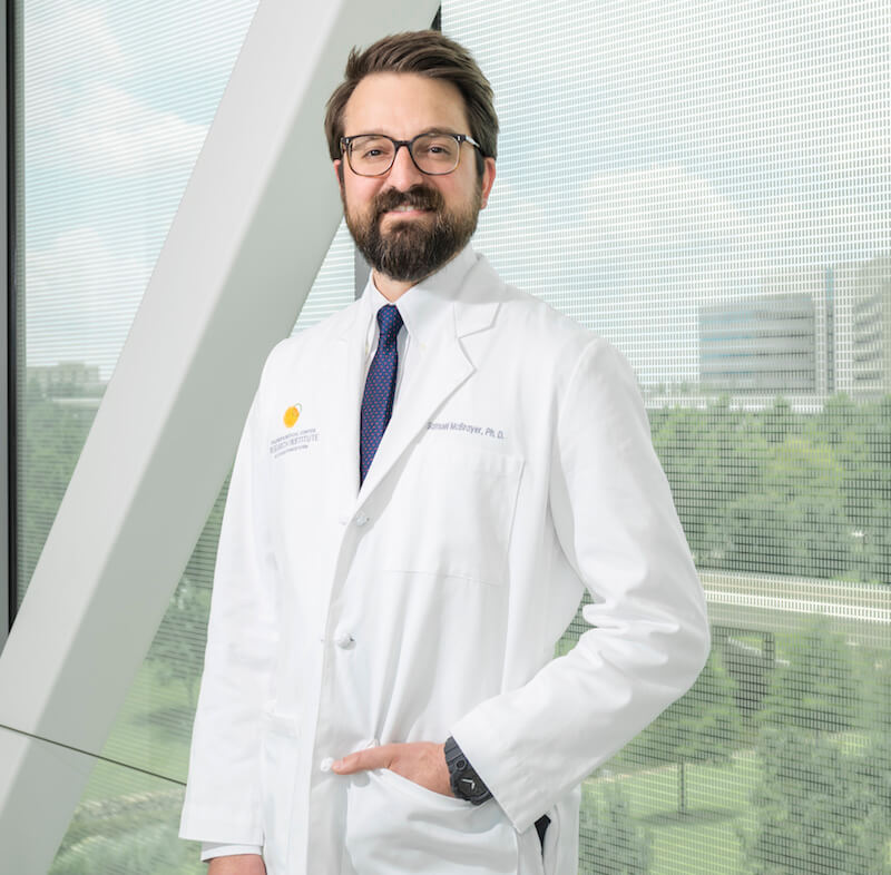
Samuel McBrayer, Ph.D.
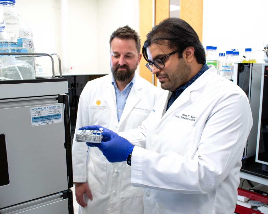
To create the organoids, researchers in the McBrayer lab worked closely with a team of physicians and scientists at UT Southwestern and with Kalil G. Abdullah, M.D., at the University of Pittsburgh. Dr. McBrayer credits the integrated cancer and research infrastructure at UT Southwestern for the project’s success.
“Very few centers have the capacity to perform this type of research. Close collaboration between the Department of Neurological Surgery and CRI and support from the O’Donnell Brain Institute and the Simmons Comprehensive Cancer Center allowed us to connect the operating room directly to the lab and collect and process brain tumor samples quickly to perform this time-sensitive research. Together, we were able to produce this breakthrough in brain tumor modeling that would not have been possible if basic scientists and clinical scientists were working alone,” Dr. McBrayer said.
The study was funded by the National Institutes of Health (R01CA258586 to Dr. Abdullah and Dr. McBrayer) and the Oligo Nation Foundation. Dr. McBrayer is a V Foundation Abeloff V Scholar, recipient of the Sontag Foundation Distinguished Scientist Award, and a Cancer Prevention and Research Institute of Texas Scholar in Cancer Research.